CONFOCAL IMAGE OF THE MONTH
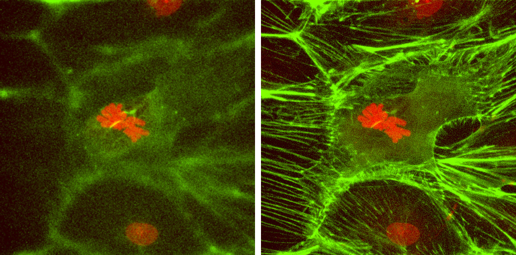
Images labeled with facpia are Ptk2 rat kangaroo endothelial
cells stained with fluorescein phalloidin (to stain actin filaments) and
propidium
iodide (to stain DNA and chromosomes). PtK cells
are tissue culture cells used in the study of mitosis because they do not
round up, but
remain relatively flat in tissue culture when undergoing
mitosis. First image: metaphase spindle showing one section through the
middle
of the spindle showing actin filaments associated with
chromosomes. Second image: same spindle, but with a second fluorescein
section
approximately 5 mm below the
first added to the image to show the actin filaments in the cells near
the substratum. Dr. Matthew J. Schibler,
Carol Moss Spivak Cell Imaging Facility, UCLA Brain Research
Institute.
|
|