 (Redrawn
from Gray)*
(Redrawn
from Gray)*
In the modern research microscopes made in the past ten years, another improvement has been made in the lens system of the compound microscope. In these microscopes, the objective lens is made to project its image at an infinite distance, hence the name infinity-corrected optics (infinity color-corrected system, ICS). In these microscopes, a tube lens is added to support the objective; it forms the intermediate image for ocular magnification. This means that after the objective lens, the light rays are all parallel until they reach the tube lens. This allows one to place other optical components (fluorescence filters, dichroic mirrors, polarizers) without disturbing the light path. That means that there is no need to add even more optics to correct for aberrations in light path that such components could introduce.
The primary image-forming component of the compound microscope is the objective lens. Knowledge about objective lenses is crucial to selecting the proper one for the microscopic technique being used and the particular specimen being observed. Please CLICK HERE for detailed discussion on objective lenses.
The most important consideration for image formation with the objective lens other than its magnification or power is its numerical aperture. This is a number which is directly related to the resolving power of the objective. It is a critical aspect in obtaining a useful microscopic image. For a discussion of numerical aperture and image resolution, CLICK HERE.
(Redrawn
from Gray)*
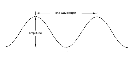
Most of the objects that biological microscopists observe are transparent; therefore there very little innate change in light amplitude or wavelength in a biological specimen observable to us. These properties are however, taken advantage of by creating changes in the light passing through the specimen. We can stain the specimen and see it in a different color than the surrounding microscopic field. This is the simplest approach because one does not need to change much about the light path in the microscope, at least for transmitted light microscopy. Some techniques which depend on specimen staining are brightfield and fluorescence microscopy. We can also alter the light path by some tricks to change the amplitude of the light passing through the specimen relative to the surrounding field. These amplitude differences created in the specimen then allow us to see it much better. Techniques which use this approach are phase, darkfield, polarization, reflection contrast, and different types of interference microscopy. Please see the discussion on phase microscopy for a further explanation of amplitude.
We have endeavored to provide some basic information about light microscopic principles in these pages for your benefit in using the microscopes and associated instrumentation in the facility. The techniques mentioned above are discussed in more detail in the following pages. Much more information is available and if you have any questions about or suggestions for this webpage, please contact the Webmaster. Below we have listed a variety of useful microscopy Web links to assist you in your work.
*Diagram redrawn from Gray, P. 1964. Handbook of
Basic
Microtechnique. McGraw-Hill: New York.
Here are some the webpages of some of the manufacturers of research
microscopes.
In the U.S.:
International:
Leica
The following links are microscopy resource web pages:
WWW Directory of Microscopy and Microanalysis Products and Services, and Vendors
Edu Sites for Microscopy and Microanalysis
The WWW Virtual Library: Microscopy